
A pelvic ultrasound is a noninvasive diagnostic pelvic exam that produces images that are used to assess organs and structures within the female pelvis.
Pelvic Sonogram
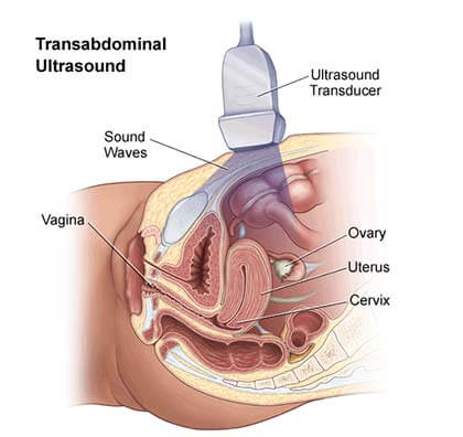 A pelvic ultrasound allows quick visualization of the female pelvic organs and structures, including:
- Uterus
- Cervix
- Vagina
- Fallopian tubes
- Ovaries
Ultrasound uses a transducer that sends out ultrasound waves at a frequency too high to be heard. The ultrasound transducer is placed on the skin, and the ultrasound waves move through the body to the organs and structures within.
The sound waves bounce off the organs like an echo and return to the transducer. The transducer processes the reflected waves, which are then converted by a computer into an image of the organs or tissues being examined.
The sound waves travel at different speeds depending on the type of tissue encountered – fastest through bone tissue and slowest through the air.
The speed at which the sound waves are returned to the transducer, as well as how much of the sound wave returns, is translated by the transducer as different types of tissue.
A Pelvic Sonogram/Ultrasound may be done to:
- Find out what is causing pelvic pain
- Look for the cause of vaginal bleeding
- Look for pelvic inflammatory disease (PID)
- Find an intrauterine device (IUD)
- Look at the size and shape of the uterus and the thickness of the uterine lining
- Look at the size and shape of the ovaries
- Check the condition and size of the ovaries during treatment for infertility
- Confirm a pregnancy and whether it is in the uterus. Pelvic ultrasound may be used early in pregnancy to check the age of the pregnancy or to find a tubal pregnancy (ectopic pregnancy) or multiple pregnancies
- Check the cervical length in a pregnant woman at risk for preterm labor
- Check a lump found during a pelvic examination
- Check uterine fibroids found during a pelvic examination. Pelvic ultrasound may also be done to check the growth of uterine fibroids
- Guide a procedure to remove an ovarian follicle for in vitro fertilization.
Other Terms that are used to describe this procedure:
- Pelvic Ultrasound
- Pelvic Ultrasonography
- Pelvic Sonography
- Pelvic Scan
- Lower Abdomen Ultrasound
- Gynecologic Ultrasound
- Transabdominal Ultrasound
- Transvaginal Ultrasound
- Endovaginal Ultrasound
Pelvic Ultrasound FAQ
How Do I Prepare for a Pelvic Ultrasound?
Normally, your healthcare provider will provide you with recommendations on how to prepare for an ultrasound procedure. Generally, it is advised to drink a minimum of 24 ounces of clear fluid at least one hour before the appointment. In the vast majority of cases, no sedation or fasting is required unless the vaginal ultrasound is part of another procedure that requires anesthesia. Keep in mind that based on your medical condition, your doctor might request specific preparation.
What Happens After a Pelvic Ultrasound?
Once your vaginal ultrasound is completed, you will be able to resume your regular diet and activity unless your healthcare provider advises you differently. Keep in mind that there is no special care required after an ultrasound procedure. Furthermore, there are no confirmed adverse biological effects on patients caused by exposure to ultrasound.
What Are the Main Goals of Pelvic Ultrasound?
This procedure is normally performed for the purpose of:
- Detecting issues with the structure of your uterus or ovaries
- Looking for cancer in your ovaries, uterus, or bladder
- Looking for growths like noncancerous tumors, fibroids, or cysts
- Discovering the cause of pain or abnormal bleeding
- Monitoring your baby’s growth during pregnancy
- Checking for pelvic inflammatory disease (PID — an infection of your uterus, ovaries, or fallopian tubes)
- Diagnosing an ectopic pregnancy (a fertilized egg that grows outside of the uterus)
What Happens During a Pelvic Ultrasound?
During a vaginal ultrasound procedure, your doctor will make use of a special device called a transducer that transmits sound waves. These sound waves bounce off your organs and tissues and then echo back to the transducer. A computer will then convert these sound waves into a picture of your organs, which will appear on a screen.
What Are the Risks of a Pelvic Ultrasound Examination?
The examination itself does not carry any risks. Unlike X-rays, vaginal ultrasound does not use radiation. Additionally, this procedure is not painful, but you might experience a certain degree of discomfort when the transducer is inserted.
Pelvic Ultrasound (Sonogram) Near Me: A Safe and Effective Diagnostic Test at Brooklyn GYN Place
Choosing a trained obstetrician-gynecologist (OB/GYN) is vital when seeking sonograms for monitoring pregnancy, diagnosing a medical problem, or looking for potential health risks since they have the skills and experience needed to conduct precise and comprehensive examinations. A qualified OB/GYN not only assures accurate results, but also provides a high level of care by explaining findings and offering vital guidance. Selecting a trained specialist who can do sonograms with proficiency and professionalism is critical to your health and peace of mind.
How to Find an OB/GYN Who Offers a Sonogram Near You
- Ask your friends, family, and other trusted sources for recommendations.
- Search online for “pelvic ultrasound near me” or “sonogram near me.”
- Call your insurance company. Many insurance companies have a list of providers in their network who offer pelvic ultrasounds.
Once you have a list of potential providers, you can start narrowing down your choices by considering the following factors:
- Is the provider’s office conveniently located for you?
- Does the provider accept your insurance?
- Is the provider experienced in performing pelvic ultrasounds?
- Does the provider offer same-day or next-day appointments?
- Does the provider have a good reputation?
We offer pelvic ultrasounds (sonograms) to women of all ages at Brooklyn GYN Place. A pelvic ultrasound is a non-invasive imaging test that creates pictures of the pelvic organs using sound waves. This can aid your doctor in the diagnosis and monitoring of a range of disorders, including ovarian cysts, uterine fibroids, and endometriosis. We also offer pelvic ultrasounds for fertility evaluation and prenatal care.
Why Choose Brooklyn GYN Place for a Pelvic Ultrasound?
- Experienced and board-certified OB/GYNs
- State-of-the-art technology and techniques
- Comfortable and supportive environment
- Personalized care
- Convenient location in downtown Brooklyn, NYC
If you are considering a pelvic ultrasound, contact Brooklyn GYN Place today to schedule a consultation. We would be happy to discuss your needs and help you determine the best course of treatment for you.
Brooklyn GYN Place
142 Joralemon Street, Suite 4CF
Brooklyn, NY 11201
(Brooklyn Heights)
718-624-0604
Would like to schedule an appointment with the top obgyn doctor in Downtown, Brooklyn, please contact our Brooklyn Heights office.
Our office has a 24/7 answering service to address
any Emergencies.
Disclaimer:
The information provided on this site is intended to educate the reader about certain medical conditions and certain possible treatment. It is not a substitute for examination, diagnosis, and medical care provided by a licensed and qualified health care professional. If you believe you, or someone you know suffers from the conditions described herein, please see your health care provider immediately. Do not attempt to treat yourself or anyone else without proper medical supervision.
- Our office has a 24/7 answering service to address any emergencies.